
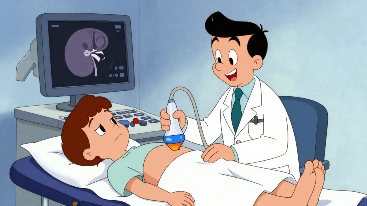
Why Your Kidneys Need a Sound Check
You feel that sharp pain in your side. It radiates down toward your groin. You might be sweating, nauseous, or just generally miserable. This is often the body’s alarm bell for a urinary tract issue. Before doctors reach for a CT scan-which involves radiation-they usually start with something simpler, safer, and surprisingly powerful: a renal ultrasound. A non-invasive imaging test using sound waves to visualize kidney structure and function..
This isn't just about taking a picture. It is about answering two critical questions quickly: Is there a blockage stopping urine from flowing? And how big are the kidneys, which tells us if they have been damaged over time? In this guide, we break down exactly what happens during this exam, what the numbers mean, and why it remains the gold standard for initial kidney evaluation.
The Basics of Renal Ultrasound Technology
Ultrasound technology has come a long way since its clinical introduction in the 1970s. Today, it is the first-line diagnostic tool recommended by major institutions like the Mayo Clinic and the Cleveland Clinic. Why? Because it uses high-frequency sound waves instead of ionizing radiation. This makes it safe for everyone, including pregnant women, children, and patients who need frequent monitoring.
The machine sends sound waves into your abdomen. These waves bounce off your internal organs and return to a probe held by the sonographer. The computer translates these echoes into real-time images on a screen. For adult patients, technicians typically use transducers with frequencies between 2-5 MHz. For smaller bodies, like children, higher frequencies (5-7.5 MHz) provide sharper detail. The entire process usually takes 15 to 30 minutes and requires no special preparation other than drinking water beforehand to fill the bladder, which helps push bowel gas out of the way.
| Modality | Radiation Exposure | Stone Detection Sensitivity | Average Cost (USD) | Best Use Case |
|---|---|---|---|---|
| Renal Ultrasound | None | ~80% for stones >3mm | $200 - $500 | Initial screening, pregnancy, serial monitoring |
| CT Urography | High (~10 mSv) | >95% (detects 1-2mm stones) | $1,500 - $2,500 | Complex cases, surgical planning |
| MRI Urography | None | Low for stones | $1,500 - $2,500 | Soft tissue characterization |
Evaluating Kidney Size and Structure
One of the primary jobs of a renal ultrasound is measuring the physical dimensions of your kidneys. Healthy adult kidneys typically measure between 9 and 13 centimeters in length. If a kidney is significantly smaller, it may indicate chronic disease or atrophy (shrinking due to lack of blood flow or long-term damage). If it is larger, it could suggest swelling from acute infection or obstruction.
Doctors also look closely at the cortical thickness, which is the outer layer of the kidney where filtration happens. A normal cortical thickness is greater than 1 centimeter. Thinning of this cortex is a red flag for chronic kidney disease. Additionally, the sonographer measures the renal pelvis anteroposterior diameter. In a healthy, non-obstructed kidney, this measurement should be less than 7 millimeters. Anything larger suggests fluid backup.
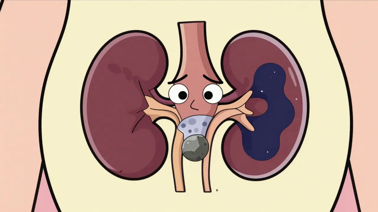
Detecting Obstruction: Hydronephrosis and Beyond
When urine cannot drain properly from the kidney to the bladder, it backs up. This condition is called hydronephrosis. The swelling of a kidney due to a build-up of urine.. On an ultrasound, this looks like dark, fluid-filled spaces expanding within the kidney structure. The severity is often graded using systems like the Society for Fetal Urology classification, ranging from mild dilation to severe, thin-walled ballooning.
Common causes of this obstruction include:
- Kidney stones (nephrolithiasis)
- Ureteropelvic junction (UPJ) obstruction, a congenital narrowing where the kidney meets the ureter
- Tumors pressing on the urinary tract
- Enlarged prostate in men
While CT scans are better at finding tiny stones (as small as 1-2 mm), ultrasound is excellent at detecting the *effect* of those stones-the swelling. According to the American College of Emergency Physicians (ACEP), point-of-care renal ultrasound has a sensitivity of 70% and specificity of 75% for diagnosing nephrolithiasis compared to CT. This means it catches most significant obstructions without exposing you to radiation.
The Resistive Index: Measuring Blood Flow
Standard ultrasound shows anatomy, but Doppler ultrasound shows physiology. By measuring blood flow through the renal arteries, doctors calculate a value called the Resistive Index (RI). A Doppler ultrasound measurement indicating vascular resistance in the kidney.. The formula is simple: (Peak Systolic Velocity minus End Diastolic Velocity) divided by Peak Systolic Velocity.
Why does this matter? When a kidney is obstructed, pressure builds up inside. This pressure squeezes the tiny blood vessels, increasing resistance. A study published in the Nigerian Journal of Clinical Practice (2015) found that an RI threshold of ≥0.70 demonstrates 86.7% sensitivity and 90% specificity for diagnosing obstructive uropathy. If your RI is above 0.70, it strongly suggests that the kidney is under stress from obstruction or inflammation.
Normal RI values are typically below 0.70. Values can vary slightly based on age and hydration status, so doctors always compare the left kidney to the right. A difference of more than 0.05 between the two kidneys is often considered abnormal and warrants further investigation.

Limitations and When Ultrasound Isn't Enough
No test is perfect. Ultrasound has specific blind spots. The biggest challenge is patient body habitus. In patients with a BMI exceeding 35, sound waves struggle to penetrate deep enough to get a clear image. Bowel gas can also interfere, blocking the view of the kidneys in 15-20% of abdominal studies.
Furthermore, ultrasound is operator-dependent. A 2018 study noted up to 20% variation in kidney length measurements between novice and experienced sonographers. This is why certification matters. The American Institute of Ultrasound in Medicine (AIUM) recommends a minimum of 40 supervised examinations for competency.
If the ultrasound is inconclusive, or if precise stone location is needed for surgery, your doctor may order a CT urography. While CT delivers about 10 mSv of radiation (equivalent to several years of natural background radiation), it provides a detailed 3D map of the urinary tract. MRI urography is another alternative, offering superior soft-tissue contrast without radiation, though it is more expensive and less effective for detecting calcified stones.
Emerging Technologies in Renal Imaging
The field is evolving rapidly. New techniques like shear-wave elastography (SWE) allow doctors to measure the stiffness of the kidney tissue. Research by Dr. S. Gennisson showed that renal stiffness increases linearly with urinary pressure. This could soon help quantify obstruction severity without relying solely on visual grading.
Another breakthrough is super-resolution ultrasound imaging. As highlighted in Nature Reviews Nephrology (2024), this technique can visualize microvascular changes in early-stage fibrosis non-invasively. Imagine detecting kidney damage before it shows up on blood tests. Additionally, AI algorithms are being developed to automate hydronephrosis grading, reducing human error and speeding up diagnosis in emergency departments.
Do I need to fast before a renal ultrasound?
No, fasting is not required. In fact, you are often encouraged to drink plenty of water beforehand. A full bladder acts as an acoustic window, pushing bowel loops away from the kidneys and providing a clearer image. However, follow your specific doctor's instructions, as protocols can vary slightly.
Can renal ultrasound detect all kidney stones?
Not all of them. Ultrasound is very good at finding large stones (>3mm) and stones causing significant swelling. However, it misses many small stones located in the mid-ureter because bowel gas blocks the view. CT scans are far more sensitive for detecting small stones.
Is a high Resistive Index always bad?
An elevated RI (≥0.70) indicates increased vascular resistance, which is common in obstruction, inflammation, or transplant rejection. However, it is not specific to one disease. Age, dehydration, and heart failure can also affect RI. Doctors interpret it alongside other clinical findings.
How accurate is point-of-care ultrasound in the ER?
It is highly useful for rapid triage. While it has lower sensitivity (70%) than CT for stone detection, it effectively rules out hydronephrosis in many cases. It reduces time-to-diagnosis by approximately 45 minutes and avoids unnecessary radiation exposure for patients with negative results.
What is the normal size of an adult kidney?
Normal adult kidney length ranges from 9 to 13 centimeters. The left kidney is often slightly longer than the right. Cortical thickness should be greater than 1 cm. Significant deviations from these norms can indicate chronic disease or acute swelling.